
Technology Advances
Cell IDx science news of new applications, new panels, and advances in our technologies.
2022
Leica Biosystems Acquires Cell IDx, Moving its Multiplexing Menu Forward with Ready-to-Use, Pre-Kitted Formulations
November 8, 2022
Leica Biosystems, a technology leader in automated staining and brightfield and fluorescent imaging, has acquired Cell IDx, Inc., a leader in multiplexed tissue profiling. Founded in 2012 and headquartered in San Diego, California, Cell IDx provides multiplex staining panels, tissue staining, and imaging and analysis services.

Cell IDx’s UltraPlex technology allows simultaneous detection of multiple biomarkers in many immunoassays through fluorescent and chromogenic multiplex immunohistochemistry (“IHC”) staining technology, and automated chromogenic detection of multiple markers on Leica Biosystems’ BOND RX fully automated research stainer. Together, UltraPlex and BOND RX with high throughput imaging allows researchers to detect three or more markers chromogenically on a single slide using a straightforward pathology workflow.
In 2021, Leica Biosystems and Cell IDx announced a partnership to combine the benefits of UltraPlex and BOND RX; now, Leica Biosystems has acquired Cell IDx, advancing this partnership and adding both scale-up capability and Medical and Scientific Affairs support for Cell IDx customers.
“We are thrilled to welcome Cell IDx to the Leica Biosystems team,” said Gustavo Perez-Fernandez, President of Leica Biosystems. “This acquisition will expand our capabilities in the translational research segment and help researchers to accelerate their journey, transforming scientific exploration into translational outcomes.”
“The ability to differentiate multiple targets in brightfield has been a long-standing unmet need in clinical pathology. Multiplex chromogenic IHC allows the pathologist to, for example, detect targets inside and outside the tumor. This data on a single tissue section has the potential to inform real time therapy decisions and produce a more focused patient centric approach,” said David Schwartz, co-founder and former CEO of Cell IDx. “Getting this technology into the hands of researchers will advance the science of cancer diagnostics and improve lives. We are delighted to be a part of this mission.”
Robert Monroe, Chief Medical Officer at Leica Biosystems, agreed with Schwartz on the value of UltraPlex. “The Cell IDx technology promotes the translation of complex multiplex IHC signatures into manageable three and four biomarker panels that can be automated on Leica Biosystems’ BOND RX platform for high quality, reproducible staining,” he said. “We see the pairing of UltraPlex and BOND automation as a platform for the creation of next generation tissue-based diagnostics for better prediction of therapeutic response and tumor behavior.”
UltraPlex fluorescent and chromogenic multiplex IHC technologies are for Research Use Only. Not for Diagnostic or Therapeutic use. BOND RX is for Research Use Only. Not for Diagnostic or Therapeutic use.
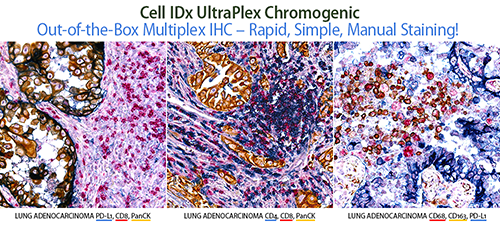
New Chromogenic Multiplex IHC Kits for Rapid Manual Staining
February 21, 2022
Cell IDx announces the release of our new chromogenic multiplex IHC kits for manual staining of human FFPE tissue sections. This simple, rapid, out-of-the-box solution allows you to stain, image and analyze, all in one day, and is an excellent complement to our current rapid BOND RX-optimized Chromogenic Multiplex IHC Kits. We invite you to view our gallery and contact us at [email protected] about custom chromogenic multiplex IHC panels!
Virtual Workshop: ZEISS Multiplex Imaging Solutions Workshop
February 8–10, 2022
Cell IDx participated in Zeiss’ workshop. Watch the presentation titled Multiplex Immunofluorescence and Multiplex Immunochromogenic Staining Employing Cell IDx’s UltraPlex Tag-based Technology and following Q&A from the workshop presented by David Schwartz, Cell IDx’s Co-Founder, Chief Executive Officer, and Chief Scientific Officer.

2021
Cell IDx Announces the release of UltraPlex Mouse Fluorescent Multiplex IHC Panels
October 22, 2021
 Announcing the
release of Mouse Fluorescent Multiplex IHC staining panels for preclinical research, the newest member of our UltraPlex technology family! Our modular format easily allows customers to build their own multiplex panel by selecting from our portfolio of biomarkers and choosing their
preferred set of fluors. Staining with UltraPlex technology either manually or on the autostainer is quick and easy, using a single antigen retrieval and two staining steps using cocktails of antibodies. Contact us at [email protected]
about additional novel biomarkers or custom chromogenic multiplex IHC panels! Read more
Announcing the
release of Mouse Fluorescent Multiplex IHC staining panels for preclinical research, the newest member of our UltraPlex technology family! Our modular format easily allows customers to build their own multiplex panel by selecting from our portfolio of biomarkers and choosing their
preferred set of fluors. Staining with UltraPlex technology either manually or on the autostainer is quick and easy, using a single antigen retrieval and two staining steps using cocktails of antibodies. Contact us at [email protected]
about additional novel biomarkers or custom chromogenic multiplex IHC panels! Read more
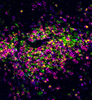
Simultaneous 6-color, 5-biomarker Multiplex Tissue Staining, with A Single Antigen Retrieval Step and No Spectral Unmixing!
June 21, 2021
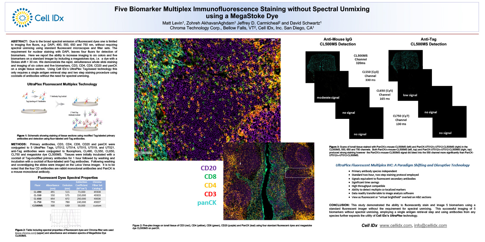 Learn how UltraPlex technology with the inclusion of a
megastokes dye can expand your biomarker profiling and enable rapid, high-throughput, 5-biomarker tissue staining and analysis using standard equipment, without the need for spectral unmixing. Download our UltraPlex poster to see how.
Read poster.
Learn how UltraPlex technology with the inclusion of a
megastokes dye can expand your biomarker profiling and enable rapid, high-throughput, 5-biomarker tissue staining and analysis using standard equipment, without the need for spectral unmixing. Download our UltraPlex poster to see how.
Read poster.
Application of Chromogenic Multiplex IHC to Lung Cancer Biomarker Profiling
April 14, 2021
Please feel free to download the poster LB238, entitled “Multiplex immunohistochemistry profiling with UltraPlex IHC on FFPE lung cancer provides a fast, robust staining platform compatible with pathology laboratory workflows,” which was presented this month at AACR21 and demonstrates the easy application of the UltraPlex technology. Thank you for stopping by our virtual booth and we look forward to seeing you in May!
2020
Did you know that Cell IDx services are now on scientist.com?
September 2, 2020
Cell IDx is a Registered Supplier with scientist.com, so sign in and send us a request today. Access our full-service multiplex biomarker tissue section staining, imaging, and analysis services using both fluorescent and chromogenic multiplex panels in human and in mouse.
Cell IDx is pleased to announce the co-authorship and use of our UltraPlex mxIF technology for profiling of the TME in this important study
March 6, 2020
 A Phase I trial assessing the safety and tolerability of a therapeutic DNA vaccination against HPV16 and
HPV18 E6/E7 oncogenes after chemoradiation for cervical cancer. View a summary of this paper by Hasan et al.
A Phase I trial assessing the safety and tolerability of a therapeutic DNA vaccination against HPV16 and
HPV18 E6/E7 oncogenes after chemoradiation for cervical cancer. View a summary of this paper by Hasan et al.
Nature: Single-cell Analysis Reveals New Evolutionary Complexity in Uveal Melanoma
January 24, 2020
Cell IDx is pleased to announce the co-authorship and inclusion of our UltraPlex mxIF technology in the University of Miami’s Nature Communications paper “Single-cell analysis reveals new evolutionary complexity in uveal melanoma.” Read the full paper on Nature’s website.
2018
Cell IDx Webinar on “Immunofluorescence Profiling of Tumor Biopsies Using UltraPlex mxIF Technology”
April 6, 2018
Please join Helen Snyder, our Director of Preclinical Development & Strategic Partnerships for a joint Cell IDx/Leica webinar looking at the application of multiplex immunofluorescence for single slide, comprehensive analysis of immune infiltrates within tumor biopsies. Watch On Demand after April 6.
GEN Feature Articles
Supplement: Who Gets Immunotherapy for Cancer?
April 1, 2018
![]() Single-agent or combination checkpoint inhibition (CPI) immunotherapy can have dramatic impacts for positive responders. But currently-used nonspecific biomarkers are unable to definitively stratify and identify cohorts. This is a critical
challenge; no one wants to deny care. Targeted biomarkers and companion diagnostics can greatly aid therapeutic decision making. Immunotherapy applications require more than just quantification of infiltrating immune cell subpopulations. Context is crucial, and concentrated efforts are
underway to provide accurate information on the percentage and location of infiltrating immune cell subpopulations, both relative to each other and to tumor margins, as well as levels of expression of checkpoint and other immune markers. The Cell IDx UltraPlex mxIF (multiplex
immunofluorescence) technology, a simple two-hour staining procedure, can simultaneously detect multiple biomarkers in tissue using a standard two-step primary and secondary antibody protocol and existing autostainers, imaging scanners, and image-analysis software. The technology is
modular (plug and play); approximately 30 primary antibodies have been qualified thus far. Read the full article from GEN.
Single-agent or combination checkpoint inhibition (CPI) immunotherapy can have dramatic impacts for positive responders. But currently-used nonspecific biomarkers are unable to definitively stratify and identify cohorts. This is a critical
challenge; no one wants to deny care. Targeted biomarkers and companion diagnostics can greatly aid therapeutic decision making. Immunotherapy applications require more than just quantification of infiltrating immune cell subpopulations. Context is crucial, and concentrated efforts are
underway to provide accurate information on the percentage and location of infiltrating immune cell subpopulations, both relative to each other and to tumor margins, as well as levels of expression of checkpoint and other immune markers. The Cell IDx UltraPlex mxIF (multiplex
immunofluorescence) technology, a simple two-hour staining procedure, can simultaneously detect multiple biomarkers in tissue using a standard two-step primary and secondary antibody protocol and existing autostainers, imaging scanners, and image-analysis software. The technology is
modular (plug and play); approximately 30 primary antibodies have been qualified thus far. Read the full article from GEN.
2017
Cell IDx Announces Publication of a Book Chapter Demonstrating the Use of UltraPlex Technology in ICC (Immunocytochemistry)
April 7, 2017
Cell IDx in collaboration with Bio-Techne announces the publication of a chapter titled “Hapten-Anti-Hapten Technique for Two-Color IHC Detection of Phosphorylated EGFR and H2AX Using Primary Antibodies Raised in the Same Host Species” in Signal Transduction Immunohistochemistry, Volume 1554 of Methods of Molecular Biology. (PUBMED). The work demonstrates the simultaneous detection of EGFR and H2AX in A431 human epidermoid carcinoma cells using Cell IDx’s proprietary hapten/high affinity anti-hapten antibody technology. This work further expands the utility of the Cell IDx UltraPlex technology for use in ICC. Further work expanding the number of biomarkers simultaneously detected in a single assay is planned.
2015
UltraPlex technology 3–4X brighter than fluorescent primaries!
June 25, 2015
Cell IDx’s multiplex immunofluorescence, breakthrough technology has solved the problem that has been perplexing histologists for decades — the ability to detect multiple markers on a single tissue!
The left panel consists of directly labeled anti-ER (blue), PR (green) and HER2 (red) antibodies. The right panel consists of anti-ER (blue), PR (green) and HER2 (red) antibodies detected using UltraPlex reagents.
Cell IDx’s UltraPlex technology provides the freedom to create multiplex IHC panels with vivid staining and without antibody species restriction. See our Multiplex Gallery.
Have you ever imagined how different your research capabilities would be if you could get multiple marker, flow-cytometry quality data on cells while also identifying their localization in tissue sections? Cell IDx has the solution!
Multiplex IHC has, in the past, been mired in difficulties:
- Fluorescent-labeled primaries just don’t give the intensity of staining that you need for sensitive detection.
- The need to use fluorescent-labeled anti-species secondaries to detect your primary antibody limits the number of markers that can be analyzed at one time.
- Attempts to solve this problem have involved repeated rounds of staining and stripping that are time consuming and can affect levels of detection.
As shown above, UltraPlex technology provides the same flexibility to detect markers that you would get using primary antibodies but provides 3–4X brighter fluorescence.
Contact us for more information.
Comparison of Cell IDx UltraPolymer Secondary Antibody-HRP Polymers versus Competitors
March 28, 2015
High sensitivity and specificity are the hallmarks of any immunodetection reagent. The development of secondary antibody-HRP polymers was a major step forward to increase sensitivity and specificity of IHC immunoperoxidase-based staining. These reagents also reduced the number of steps and significantly shortened the time required to stain tissues. However commercially available secondary antibody-HRP polymers produce widely disparate images with respect to sensitivity and specificity.
Cell IDx introduces our UltraPolymer Secondary Antibody-HRP Polymers that produce images with equal or greater sensitivity and specificity compared to clinically used polymers. The image below compares Cell IDx anti-mouse and anti-rabbit secondary antibody-HRP polymers to both Dako and Biocare’s polymers. If you are interested in comparing our UltraPolymers to the HRP polymer you are currently using, please let us know.
We welcome any suggestions for other high sensitivity peroxidase reagents to evaluate by comparison. We want to prove that we offer the most sensitive, most specific, and most cost-effective peroxidase conjugates you can buy!
Staining of ER+ PR+ Ki-67+ breast carcinoma sections using anti-ER and anti-PR rabbit monoclonal antibodies and anti-Ki-67 mouse monoclonal antibody and detecting using either Cell-IDx’s UltraPolymer Secondary Antibody-HRP polymers or Biocare’s Mach4 or Dako’s Envision+ staining systems. Click for more detailed staining protocol and image.
Page Contents
Cell IDx is a Registered Supplier on
![]() and
and
![]()
© 2016–23 Cell IDx. Cell IDx, UltraPlex, UltraPolymer, UltraTag, and Path IDx are trademarks of Cell IDx, Inc. All Rights Reserved. Privacy Policy | Terms of Service Website by Fifth Street Design.